Before we begin to talk about gene therapy, or if you just need to review the basics, let’s begin at the cellular and molecular level to see what exactly genetics is and how it really works. The Bare Bones of Genetics As you may know, all living organisms have certain characteristics or traits, features which distinguish them. Your hair color is a trait. Your eye color is also a trait. Many organisms have similar traits; some organisms have different traits. It’s a difference in these traits that gives us the diverse variety of organisms prevalent on Earth today. So, where do these traits come from? Well, what your cells do (to make those traits) is dictated by the commands they receive. These commands basically come from DNA and the proteins it eventually indirectly makes. DNA is the short form of deoxyribonucleic acid. You have DNA in each and all of your cells: lots of it! DNA is packed together and coiled (very tightly!) in bundles around histones, which are proteins. Histones are like the tape roll holder in a tape role: the tape (DNA) is wrapped around the holder (histone). A certain amount of this DNA, histone and other proteins forms chromatin. A large amount of this supercoiled chromatin condenses together and forms a chromosome. Each organism has a different number of chromosomes. Humans have 46 chromosomes in total. 44 of these chromosomes are autosomes. 2 of these chromosomes are sex chromosomes. The sex chromosomes dictate gender: females have two X chromosomes, and males have one X and one Y chromosome. Now, let’s go back to the DNA.  A three-dimensional molecular representation of DNA. What secrets does this complex yet simple molecule of life hold?  A representation of DNA, labeled. Notice how the base pairs are complementary: A pairs with T, and C pairs with G.
 Replication, Transcription, & Translation
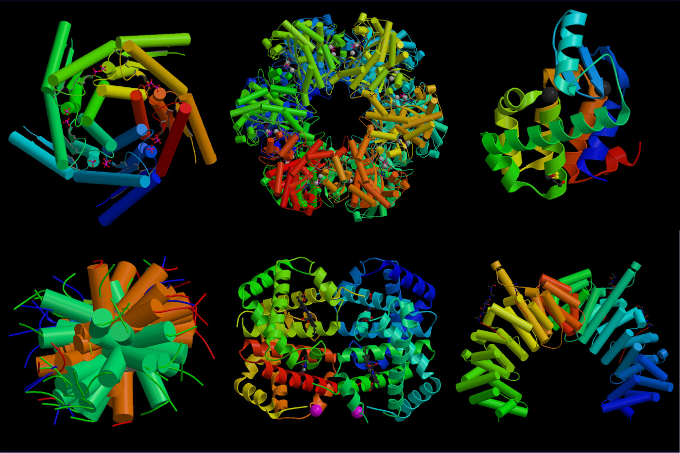
Replication Replication starts when the DNA helix is unwound by an enzyme called helicase. Helicase disrupts the hydrogen bonds and exposes the DNA base codes/nucleotides. Now that the double helix of DNA is two separate strands, each strand will function as a template to synthesize a new double strand of DNA. DNA replication then occurs on both strands, with the enzyme DNA polymerase running across both strands and forming new strands. Thus, DNA polymerase functions to add bases to the new DNA chain. It runs across the strand and swaps each nucleotide with a complementary nucleotide: A is replaced with T (and vice versa) and C is replaced with G (and vice versa). DNA polymerase also proofreads the chain for mistakes afterward. Do you remember how one strand of the original DNA ran in the 5’ to 3’ direction, whereas the other ran from 3’ to 5’ direction (antiparallel arrangement)? Well, DNA polymerase is correctly oriented for synthesis only in the 5’ to 3’ direction strand, called the leading strand. This strand can be synthesized as a continuous and complete strand. The other strand, however, called the lagging strand, runs the other way (3’ to 5’). Thus, DNA polymerase must make a copy of this strand backwards in short sections. These sections are known as Okazaki fragments, after the scientists who discovered them, Reiji and Tsuneko Okazaki. Later, these sections are joined together to form one continuous strand by an enzyme called DNA ligase.  The various types of RNA. RNA has ribose as a sugar, instead of deoxyribose like DNA. (Image Citation 11) A general diagram of transcription. Notice that there is no thymine in the RNA strand. Instead, uracil replaces all adenines. The process of translation. The polypeptide chain is a chain of amino acids. The ribosome consists of two units; namely, the large subunit and the small subunit.  What's happening inside the ribosome when translation occurs?  The beautiful structures of proteins; the folding allows the proteins to carry out their functions with great efficiency. (Image Citation 19)  |
 A view of deoxyribonucleic acid (DNA) molecule  DNA is tightly coiled around proteins called histones. Histones, in turn form chromatin, which forms chromosomes. The Structure of DNA The structure of DNA was first described by James Watson and Francis Crick. DNA is a double helix: this means that it has two sides/strands twisted around each other. Connecting both of these sides are varying types of nucleotides, as well as hydrogen bonds that connect these bases. A nucleotide consists of a nitrogenous compound, a phosphate, and deoxyribose, the sugar that gives DNA its name. There are four types of nucleotides in DNA: Adenine (A), thymine (T), guanine (G), and cytosine (C). These, as mentioned before, are located on both sides of the DNA and are bound by hydrogen bonds. Thus, one nucleotide connected to another nucleotide by a hydrogen bond forms a “rung” on the ladder-like DNA structure. Now, there are rules which govern what nucleotides are connected to what nucleotides by the hydrogen bonds. Adenine (A) always pairs with thymine (T), and vice versa. Similarly, guanine (G) always pairs with cytosine (C), and vice versa. One side of the DNA runs in a 5’ to 3’ (the ‘ sign means “prime”) direction, and the other in the 3’ to 5’ direction. This is called an antiparallel arrangement – Both strands of DNA thus run in opposite directions. In eukaryotic organisms, such as humans, DNA is located inside the nucleus of the cell. DNA serves to synthesize certain proteins. A set of DNA which codes for a protein or a trait is known as a gene. According to Talaro’s “Foundations in Microbiology: Fourth Edition”, “Genes fall into three basic categories: structural genes that code for proteins, genes that code for RNA [ribonucleic acid], and regulatory genes that code for gene expression” (Citation 36). All of the organism’s genetic makeup forms what is known as the genotype of the organism. When the genotype is expressed, traits are created, and these are called the phenotype. For example, a set of genes (genotype) codes for your hair color, which is your phenotype. The proteins are the building blocks of life: they tell the cell what to do, they tell other cells what to do, they make the structure of the cell, and much more. Well, how does the DNA make these proteins? To do so, it has to first replicate, and then it must go through the processes of transcription and translation. The processes of replication, transcription and translation always take place when a cell divides. This is to give a copy of the chromosome to the cells produced. Translation also allows proteins to be produced.  A look at DNA replication, with the inset showing a larger and general view. "Pol" stands for polymerase, a key enzyme. Note how each enzyme works in a 'biochemical team' to complete the process efficiently.  General diagram of the steps involved in DNA replication. This is how the DNA replicates to form additional chromosome during cell division. Transcription DNA does not perform cell processes directly by itself. Rather, it is converted to RNA (ribonucleic acid) first. Therefore, once replication has occurred, transcription takes place. DNA is copied onto RNA by the process of transcription. In transcription, RNA is formed by DNA by mounting nucleotides onto an mRNA strand (messenger RNA). Transcription begins when an enzyme called RNA polymerase recognizes a specific segment of the DNA which is called the promoter region, which is usually a palindrome-sequence of nucleotides. As the mRNA is made by adding nucleotides, the DNA starts to wind back into its original double helix form. RNA, unlike DNA, is a single stranded molecule, not a double helix. Also, RNA carries the sugar ribose, not deoxyribose. mRNA is messenger RNA. It will play an important role in the synthesis of the protein it is coding for. Once again, the nucleotides added are complementary to each other. However, there is a difference. There is no thymine in the mRNA or in RNA in general. Instead, adenine pairs with a nucleotide called uracil. Therefore, any T nucleotides turn into A. Any A nucleotides turn unto U. Any G nucleotides turn into C, and any C nucleotides turn into G. The nucleotides being mounted onto the mRNA from the DNA form a series of triplets, each triplet consisting of three nucleotides respectively. Each triplet is known as a codon. The DNA template from which nucleotides are being mounted is known as the template strand. Translation Once transcription has been completed, the mRNA travels out of the nucleus of the cell into the cytoplasm to one of the many ribosomes for translation. A ribosome is like a mobile factory in the cell, which serves to translate the mRNA. The mRNA goes into the ribosome, and codon by codon, it is synthesized. Synthesis begins with a start codon, which is mostly AUG. When AUG enters the ribosome, a tRNA molecule enters the ‘P site’ on the ribosome. tRNA is transfer RNA. It is a t-shaped molecule on one end. This end carries an anticodon, which is simply the complementary triplet to the mRNA codon which it is bringing an amino acid for. For example, the anticodon for AUG would be UAC. The other end of the tRNA carries an amino acid. A special note on amino acids: they are the building blocks of proteins themselves. There are 20 different types of amino acids. Each codon on the mRNA codes for a specific one of those 20 amino acids; for example, AUG codes for formyl methionine. But, there is also a factor in existence known as degeneracy: this refers to the fact that sometimes, more than one type of codon can code for a specific amino acid. Now, the tRNA for AUG carries the amino acid formyl methionine. Once the tRNA is in place, another tRNA enters the A site. It carries the amino acid for the next codon. Note that the initial tRNA was still in position. The amino acids from both of the tRNAs form a dipeptide bond. Then, the ribosome translocates, which means that it moves to the right of the mRNA. This causes the now blank tRNA to discharge from the ribosome from the E site. The tRNA holding the dipeptide is shifted off to the P position. Site A is left empty and can now hold another tRNA to code for the next codon. The blank tRNA that was discharged drifts off and can be reused later for other translations. At the ribosome, tRNA molecule number 3 enters and brings another amino acid. It binds to the correct codon at the now-empty site A. Its amino acid forms a bond with the dipeptide, making a tripeptide. tRNA molecule number 2 is now empty, is discharged, and shifts the mRNA to the next position. This moves tRNA 3 to position P, leaving position A open for the next molecule. Thus, the mRNA moves along the ribosome, being coded for by tRNA molecules carrying the correct amino acid. As mentioned before, each tRNA molecule has an anticodon which binds to the ribosome when its complementary codon on the mRNA passes through. The end of the mRNA is marked by nonsense codons: they have no corresponding tRNA molecules (and thus no amino acids) but they serve an important function by telling the ribosome to stop synthesis. Remember the chain of amino acids that was forming at the top of the ribosome? Well, once all of the mRNA has been synthesized, the chain breaks off by the use of special enzymes. It is now a chain of amino acids: a protein. As you may know, mRNA carries hundreds or thousands of codons. While the translation was occurring, the chain of amino acids began to fold itself into a complex structure. This process is called protein folding and it’s important because the shape of proteins often plays a role in their function or working.
<---An Overall Review of Replication, Transcription, and Translation
|
See the following video in order to review the concepts of transcription, translation and protein synthesis. |